Hyaline Cartilage
|
|
|
|
Hyaline cartilage is the most widely distributed cartilage in the body. It is found at the ends of long bones (articular cartilage),within joints (menisci), makes up the embryonic skeleton, supports the nose, in the trachea and larynx and it is the cartilage of the costal cartilages. It protects the ends of bone from wear and tear, it supports structures like the trachea and nose. You have probably seen hyaline cartilage while enjoying your favorite meal of fried chicken. Hyaline cartilage is the shiny white tissue on the ends of the chicken leg bones; it is also the white tissue at the tip of the chicken breast bone.
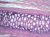
The matrix is composed of collagen. The fibers are very, very fine and do not stain individually. This tissue has a very even, smooth texture punctuated by lacunae and chondrocytes when seen under the microscope.The lacunae appear to be randomly scattered around the matrix.
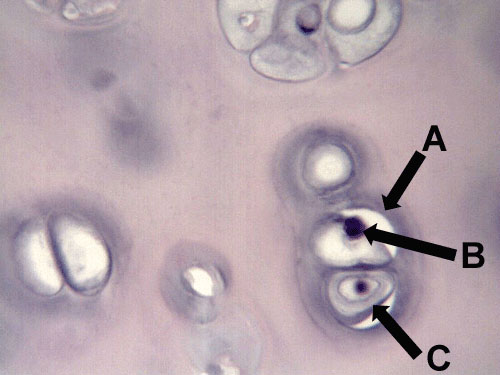
The upper image to the right is a photomicrograph of hyaline cartilage. The yellow star is on the matrix. The blue arrows are pointing to chondrocytes and the green arrow is pointing at a lacuna. The image below is a higher magnification of this same slide. Letter 'A' is pointing to the lacuna and letters 'B' and 'C' are pointing to chondrocytes.

