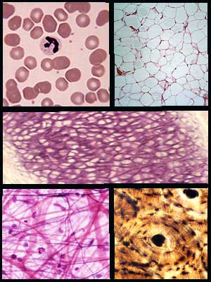
Tissues shown from left to right and top to bottom. Row 1: Blood, Adipose, Row 2: Elastic Cartilage, Row 3: Areolar, Osseous
Histology of the Connective Tissues
Review of connective tissues
|
|
|
Tissues shown from left to right and top to bottom. Row 1: Blood, Adipose, Row 2: Elastic Cartilage, Row 3: Areolar, Osseous
|
Histology is the study of tissues. A tissue, you may recall is a collection of cells that has a particular function. Histology and pathohistology (the study of disease processes in tissues) is rarely the allied health student's favorite unit in the course. Histology is unfamiliar and to the uninitiated, challenging to decipher. Hopefully, this learning module will assist you in deciphering the connective tissues. It is important. Disease and disease processes start at the level of the cell and are typically first detected by histological and physiological changes that occur at the tissue level. To understand histology, is to understand the health condition of the patient.
There are only four types of tissue that make up the human body: connective, epithelial, nervous and muscular. Together these four types of tissue make up every organ and every organ system in the body. The diversity and variety of cells and the tissues they produce is truly amazing. This module will examine connective tissues.
Connective tissue is the most common tissue found in the body. Its name describes its function. Connective tissues connect parts of the body, like muscle to bone or epithelium to the underlying tissue. The connective tissues include: osseous (bone), blood, areolar, adipose, cartilage, dense connective (regular, irregular), and loose connective. Connective tissues like bone, adipose and areolar tissue have protective and cushioning functions. The skull's protective function is pretty clear. However, adipose tissue or fat also protects and insulates organs as does areolar tissue. Additionally, fat serves as an energy storage mechanism. Connective tissues generally have a good blood supply. The cartilages are an exception; they are not well vascularized.
Students are often overwhelmed when trying to identify a particular tissue by its microscopic appearance. The slide is a riot of color and cells and seemingly with out order. Remember a slide of an organ may have representations of all of the tissue types present. In order to identify the tissue type you will need to answer some very simple questions. The first question you need to ask yourself is, 'Does this tissue have a free surface?'. If the answer is yes, then this is an epithelial tissue (discussed in another module). If the answer is no then the tissue is either connective, muscular or nervous tissue. The next question you should ask is 'Does this tissue contain matrix?'. If the answer is no, then the tissue is either muscle or nervous tissue. If the answer is yes, then this tissue is a connective tissue. We will continue the discussion of connective tissue on the next page.

The connective tissues are divided into 4 main types of tissue: blood, bone, cartilage, and connective tissue proper. The connective tissues proper category includes dense fibrous, reticular, loose, adipose and areolar tissue. Each of these tissues is characterized by the tissue's matrix and resident cells.
Connective tissue is characterized by its matrix and the types of cells associated with the tissue. The matrix is the extracellular material surrounding the cells. It is composed of ground substance and fibers. The amount of matrix and the consistency of the matrix varies with the connective tissue.The ground substance varies from a liquid consistency like the plasma of blood to a gel-like consistency found in areolar tissue to a solid matrix like that found in osseous tissue or bone. Fibers produced by the tissues' cells are also suspended in the ground substance. These fibers include reticular fibers, collagen fibers, and elastic fibers. These will be discussed later. A tissue is also characterized by the types of cells associated with the tissue. Areolar tissue contains fibroblast cells that produce the fibers found in the matrix and mast cells for immune response. Osseous tissue contains osteocytes which produce the bony matrix. Cartilage contains chondrocytes which produce the cartilage matrix.
Keep in mind that you need to be able to identify these tissues in a microscopic section. You should be able to provide a description of their function and a location in the body where the tissue would be found.
|
|
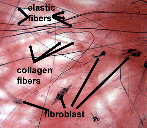
Areolar tissue is a loose connective tissue with a jelly-like matrix containing elastic, reticular and collagenous fibers. Reticular fibers are fine strands of collagenous fibers wrapped in elastic fiber. Reticular fibers are very difficult to see in areolar tissue. They are very, very fine and if visible will stain a dark color. Elastic fibers are sometimes called yellow fibers because tissue rich in elastic fibers appear yellowish in an unstained state. When stained, elastic fibers are thin, branching black lines. They give the tissue resilience so that it can stretch and rebound. Collagenous fibers are sometimes called white fibers because tissues rich in collagenous fibers in an unstained state appear white.
Cells commonly observed in this tissue include fibroblasts, mast cells, macrophages and other white blood cells.
Areolar tissue is found beneath all epithelial tissues, in the breast and surrounding organs. It cushions organs. Its location under epithelium and the presence of immune cells in this tissue allow this tissue to play a significant role in host defense.
|
|
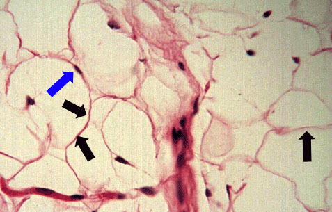
Adipose tissue is composed of cells called adipocytes. The adipocytes contain a very large fat-containing vacuole. This gives the cell the appearance of being empty. The nucleus is visible and usually appears squashed up against the plasma membrane. There is little matrix to this tissue.
Adipose tissue or fat is found just about everywhere, in your eyelids, around your organs, beneath your skin, behind your eyeballs, everywhere..... Adipose tissue plays an important role in protecting and cushioning parts of the body. It serves as an energy reserve and as a thermal insulator.
The photomicrograph to the right is adipose tissue. Notice the big empty-appearing adipocytes. The blue arrow is pointing to an adipocyte nucleus. The black arrows are pointing to the plasma membranes of the cells.
|
|
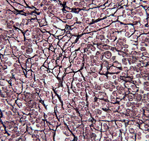
Reticular tissue is composed of a loose ground substance with distinctive reticular fibers. The reticular fibers stain black and blood cells (lymphocytes) are the predominant cell type. The reticular fibers provide a loose, flexible scaffold on which the cells rest. Reticular tissue is associated with lymphoid tissue like, lymph nodes.
In the photomicrograph to the right the reticular fibers are evident as dark black lines. The cells associated with the fibers are lymphocytes.
|
|
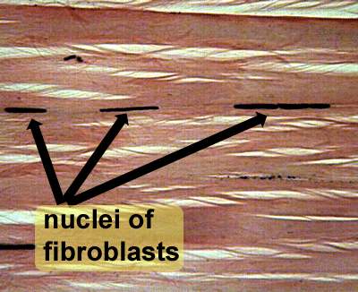
Dense regular connective tissue is composed almost exclusively of tightly packed collagen (white) fibers. There may be a few elastic fibers interspersed in the ground substance. The predominant cell type is the fibroblast. Tendons and ligaments are composed of dense connective tissue. It functions to anchor muscles to bone and bone to bone. It has to be strong, not too elastic and it must withstand tensile forces. The parallel arrangement of fibers gives this tissue a lot of strength typically in one direction.
The photomicrograph to the right is an example of dense regular connective tissue. The pink fibers are thick bands of collagen. The arrows are pointing to the nuclei of fibroblasts squeezed in between the fibers.
|
|
This dense connective tissue contains a large amount of elastic fibers. This allows the tissue to stretch and recoil. Dense regular connective tissue tends to be 'brittle' and dense elastic tissue tends to be more elastic. This tissue is associated with large arteries close to the heart that undergo routine expansion, ligaments of the vertebral column and with the air passageways of the lungs.
The photomicrograph to the right is a section from an elastic artery. Notice all of the dark blue staining elastic fibers.
This tissue is composed primarily of collagen fibers, a few elastic fibers and a few fibroblasts. The composition of dense irregular is much like dense regular; it is the arrangement of the fibers that distinguishes these two tissue types. In dense irregular connective tissue, the collagen fibers appear to be haphazardly arranged, not like the neat, parallel arrangement seen in dense regular tissue. Because the fibers extend in various directions, this tissue can withstand stress in multiple directions. This is the tissue found in joint capsules, sclera of the eye, dermis of the skin and submucosa of the digestive tract.

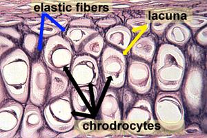
There are three types of cartilage: hyaline, elastic and fibrocartilage. All three are composed of collagen fibers, but they vary dramatically in the amount of elastic fibers present in the tissue. The tissue characteristically has spaces or chambers called lacuna (lacunae, pl.) in which the cells are encased by matrix. The mature cells found in the lacunae are called chrondrocytes. They form the matrix.
The functions, locations and characteristics of each type of cartilage will be discussed on the next few pages. Cartilages do vary in the location where each type is found in the body and in their function. However, they do not vary in the observation that cartilages are only slightly vascularized. They do not have extensive networks of blood vessels passing through the tissues. Physiologically that means nutrients have to diffuse from the blood vessels through the tissue and waste products similarly have to diffuse from the cells to the bloodstream. Clinically, this also translates into prolonged convalescence or healing time when cartilages are damaged. Bone, muscle and most other tissues heal more quickly than damaged cartilage.
|
|
|
|
Hyaline cartilage is the most widely distributed cartilage in the body. It is found at the ends of long bones (articular cartilage),within joints (menisci), makes up the embryonic skeleton, supports the nose, in the trachea and larynx and it is the cartilage of the costal cartilages. It protects the ends of bone from wear and tear, it supports structures like the trachea and nose. You have probably seen hyaline cartilage while enjoying your favorite meal of fried chicken. Hyaline cartilage is the shiny white tissue on the ends of the chicken leg bones; it is also the white tissue at the tip of the chicken breast bone.
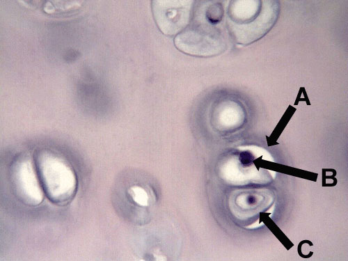
The matrix is composed of collagen. The fibers are very, very fine and do not stain individually. This tissue has a very even, smooth texture punctuated by lacunae and chondrocytes when seen under the microscope.The lacunae appear to be randomly scattered around the matrix.
The upper image to the right is a photomicrograph of hyaline cartilage. The yellow star is on the matrix. The blue arrows are pointing to chondrocytes and the green arrow is pointing at a lacuna. The image below is a higher magnification of this same slide. Letter 'A' is pointing to the lacuna and letters 'B' and 'C' are pointing to chondrocytes.
|
|
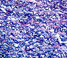
Elastic cartilage is so named because of the large amount of elastic fibers found in the tissue. Elastic fibers give the tissue elasticity and allow the tissue to recoil in response to stretching or stress. Elastic cartilage is found in the pinna of the ear and the epiglottis. Grab your ear, bend it and then release it. Your ear springs back to its original position. It does this because the elastic cartilage in the ear is resilient and recoils. If your ear contained hyaline or fibrocartilage it would not be nearly as flexible or resilient. You could actually break your ear.
Under the microscope elastic cartilage looks very different from hyaline cartilage. Chondrocytes and lacunae are still present but the matrix contains darkly staining elastic fibers. These fibers can be scattered around the tissue or they can be very densely arranged in the tissue.

|
|
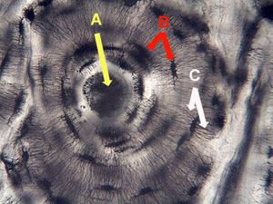
Bone or osseous tissue has a matrix composed of a solid material called apatite. The matrix is deposited by osteoblasts and osteocytes. Osteoblasts are pre-cursor cells that deposit the bony matrix. As they become encased in bone we call them osteocytes. Compact bone is organized into structures called osteons. The osteons run parallel to the length of the bone. In cross section the osteon resembles a bull's eye. At the center of the bull's eye is the osteonic or Haversian canal, a passageway for an arteriole, veinule and nerve. Surrounding this passageway are layers of bone, shells of bone called lamellae. Visible within the lamellae are dark spots or pits called lacuna. The osteocytes are found in the lacuna. Emerging from the lacunae are lines that extend from the lacuna of one lamella to the lacuna in another lamella. These lines are the canaliculi. They are actually fine grooves in the bone that allow osteocytes to pass along nutrients and waste products. When you first look at your slide the bull's eye structure is easily seen. The spider-like structures are the lacuna (spider body) and the canaliculi (spider legs).
The letter 'A' is pointing at the Haversian or osteonic canal. The arrows associated with the letter 'B' are pointing at lacuna and the arrows associated with the letter 'C' are pointing at the canaliculi.
|
|
|
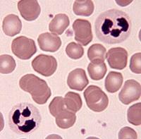
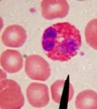
Blood is a connective tissue. Its matrix is plasma. Within the plasma are various cells (red blood cells, white blood cells), cell fragments (platelets) and dissolved macromolecules.
The images to the right are photomicrographs of a blood smear stained using Wright's stain. The two large multilobed cells in the first image to the right are neutrophils, the most common leukocyte or white blood cell. The neutrophils are surrounded by anucleate red blood cells or erythrocytes. Red blood cells are the most common cells seen in a normal blood smear. In the far right image, the large cell containing the orange granules is an eosinophil, another type of leukocyte. Eosinophils are much rarer than neutrophils. They are the leukocytes that are especially effective against parasites. The pinkish/bluish dust-like particles in both images are platelets. Cell fragments that play an important role in clotting.
Please watch the You Tube video below to review how to identify the connective tissues. Then complete the review activities on the next two pages.