Histology: Connective Tissue
|
|
|
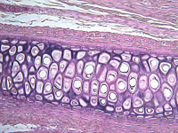
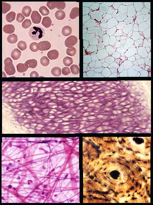
Tissues shown from left to right and top to bottom. Row 1: Blood, Adipose, Row 2: Elastic Cartilage, Row 3: Areolar, Osseous
|
Histology is the study of tissues. A tissue, you may recall is a collection of cells that has a particular function. Histology and pathohistology (the study of disease processes in tissues) is rarely the allied health student's favorite unit in the course. Histology is unfamiliar and to the uninitiated, challenging to decipher. Hopefully, this learning module will assist you in deciphering the connective tissues. It is important. Disease and disease processes start at the level of the cell and are typically first detected by histological and physiological changes that occur at the tissue level. To understand histology, is to understand the health condition of the patient.
There are only four types of tissue that make up the human body: connective, epithelial, nervous and muscular. Together these four types of tissue make up every organ and every organ system in the body. The diversity and variety of cells and the tissues they produce is truly amazing. This module will examine connective tissues.
Connective tissue is the most common tissue found in the body. Its name describes its function. Connective tissues connect parts of the body, like muscle to bone or epithelium to the underlying tissue. The connective tissues include: osseous (bone), blood, areolar, adipose, cartilage, dense connective (regular, irregular), and loose connective. Connective tissues like bone, adipose and areolar tissue have protective and cushioning functions. The skull's protective function is pretty clear. However, adipose tissue or fat also protects and insulates organs as does areolar tissue. Additionally, fat serves as an energy storage mechanism. Connective tissues generally have a good blood supply. The cartilages are an exception; they are not well vascularized.
Students are often overwhelmed when trying to identify a particular tissue by its microscopic appearance. The slide is a riot of color and cells and seemingly with out order. Remember a slide of an organ may have representations of all of the tissue types present. In order to identify the tissue type you will need to answer some very simple questions. The first question you need to ask yourself is, 'Does this tissue have a free surface?'. If the answer is yes, then this is an epithelial tissue (discussed in another module). If the answer is no then the tissue is either connective, muscular or nervous tissue. The next question you should ask is 'Does this tissue contain matrix?'. If the answer is no, then the tissue is either muscle or nervous tissue. If the answer is yes, then this tissue is a connective tissue. We will continue the discussion of connective tissue on the next page.


