Blood
|
|
|
Blood is a connective tissue. Its matrix is plasma. Within the plasma are various cells (red blood cells, white blood cells), cell fragments (platelets) and dissolved macromolecules.
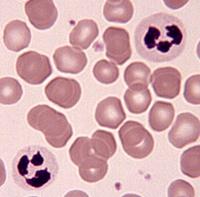
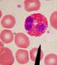
The images to the right are photomicrographs of a blood smear stained using Wright's stain. The two large multilobed cells in the first image to the right are neutrophils, the most common leukocyte or white blood cell. The neutrophils are surrounded by anucleate red blood cells or erythrocytes. Red blood cells are the most common cells seen in a normal blood smear. In the far right image, the large cell containing the orange granules is an eosinophil, another type of leukocyte. Eosinophils are much rarer than neutrophils. They are the leukocytes that are especially effective against parasites. The pinkish/bluish dust-like particles in both images are platelets. Cell fragments that play an important role in clotting.