Rat Dissection
To dissect,literally means to expose to view. It is important when doing dissections to make sure you know what you are doing before you start cutting. You do not want to hack and chop. You want to view all of the structures in place or in situ, so work carefully with your specimen. Scissors, not the scalpel are the preferred instrument particulary when you are cutting through the skin and body wall. Lift the structures away from the underlying tissues and organs with your fingers and make a small incision with your scissors. Then insert the sharp point of the scissor into the opening and cut as you lift the tissue (skin or abdominal muscle layer).
Follow the directions given in your laboratory manual.
1. Expose the abdominal muscles by first cutting through the skin. Peel the skin back and pin the skin down to your pan.
2. Carefully cut through the abdominal muscles along the midsagittal line. You can do this by lifting the muscle with forceps (tweezers) and inserting your scissors above the organs.
3. You will need to cut through the ribcage.
4. Make tranverse cuts below the diaphragm to allow you to open these cavities easily.
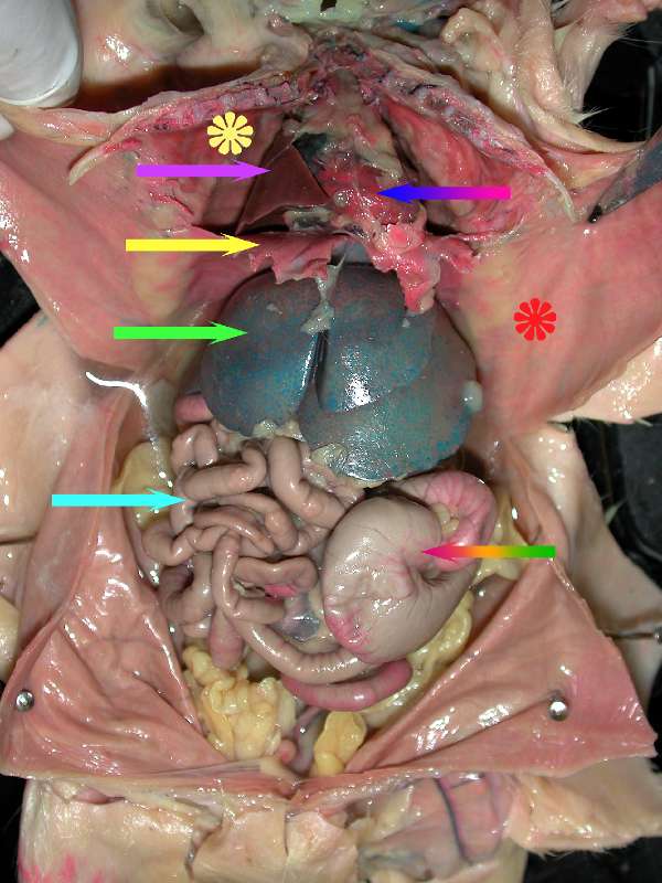
Identify the organs in the rat's thoracic and abdominal cavities. Use your lab manual as a guide. Some of the organs are shown in the photographs below.
 The diaphragm is shown at the tip of the yellow arrow. The space superior to the diaphragm is the thoracic cavity.
The solid pink arrow is pointing to a lobe of the right lung.
The yellow flower is located on the serous membrane surround the pleural cavity, ie. parietal pleura.
The blue to red rainbow arrow is touching the parietal pericardium, the sac surrounding the heart.
The thymus is visible superior to the heart in the mediastinum.
The red flower is on the serous membrane of the abdominal wall or the parietal peritoneum.
The light green arrow is pointing to a lobe of the liver, the largest abdominal organ. Notice the shiny surface of the liver and the other organs. They are shiny because the organs are encased in serous membranes called visceral serosa.
The light blue arrow is pointing to the small intestine.
The red to green rainbow arrow is pointing to the cecum of the large intestine.
