The Cell_2
Anatomy and Division
|
|
The cell is the fundamental unit of all life. Life in its simplest form is a single cell. In its most elegant form (arguably), the human body is composed of billions of cells. Whether an organism is composed of one cell or billions there are certain common functions that must be done, metabolism and reproduction just to name two. From an anatomy and physiology viewpoint understanding the cell and its functions is key to understanding normal and disease conditions.
This module will cover the basics of animal cell structure and cell somatic division.
The plasma membrane is a dynamic component of cell structure and surrounds all living cells. The plasma membrane separates the inner workings of the cell from the environment. It maintains homeostasis, the constant internal condition needed for life. The plasma membrane is composed primarily of lipids and proteins. It is a semi-permeable or selectively permeable membrane. The selective permeability of the membrane maintains homeostasis by controlling what enters and what leaves the cell. The cell membrane and pertinent transport mechanisms have been discussed in the transport and diffusion lab. Further discussion will not occur here.
The plasma membrane exhibits modifications that better equip the cell for its function. Epithelial cells of the small intestine function in nutrient absorption. Their apical surface is covered by microvilli. These microvilli are nipple-like or fingerlike projections that dramatically increase the surface area of the cell. More area means more effective absorption of nutrients. Epithelial cells of the trachea, nasal passageways and oviducts have cilia on their apical surfaces. Cilia are fine, short extensions of the membranes that move substances like mucus or the ovum past the surface of the cell.
The cytoplasm is found within the plasma membrane. It is the cell 'jelly' within which the cell organelles are suspended and cell solutes are dissolved. Metabolism occurs in the cytoplasm and in the organelles suspended within the cytoplasm. Some texts define the cytoplasm as everything inside the cell except for the nucleus. Cytosol is another term frequently used in discussions of cell anatomy. Cytosol is the liquid component of the cytoplasm, i.e., the cytoplasm without organelles.

|
|
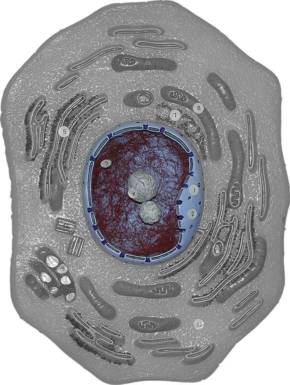
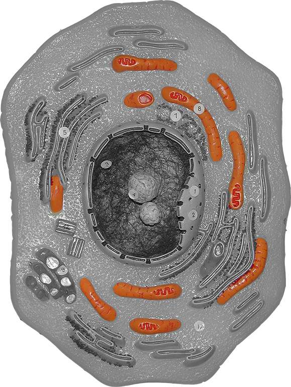
The nucleus is a large membrane-bound organelle within the cell that contains the cells DNA, or genetic material. The image to the right is an edited photograph of one of the animal cell models we have in lab. The structure highlighted in blue is the nucleus. In this cell, as in many other cells the nucleus is located in the middle of the cell.
The membrane surrounding the nucleus is called the nuclear membrane or nuclear envelop. This membrane has many six sided pores called nucleopores that allow information molecules (remember we copy information from the DNA template) to move from the nucleus to the cytoplasm.
The nucleus is filled with nucleoplasm. The nucleoplasm is jelly-like and very similar to cytoplasm in its consistency. Suspended within the nucleoplasm are the nucleoli and the chromosomes. Nucleoli (nucleolus, singular) are the sites of ribosome production. Ribosomes are the workbenches of protein synthesis and will be discussed more later. For now, it is sufficient to say that they are produced in this region of the nucleus. Within the nucleoli you would find the RNA and protein needed for the production of the ribosomes.
All of the information a cell needs to function is stored in our genetic molecules or in our DNA. Your DNA is the master set of instructions, the blueprint or the play book for making you, you. Our genetic information is organized in the form of chromosomes. Humans have 46 chromosomes. We have 23 pairs of chromosomes. Chromosomes 1-22 are called autosomes and the last pair are the the sex chromosomes. We inherit one chromosome of each pair from each of our parents. So your Dad contributed 23 chromosomes in his sperm and your Mom contributed 23 chromosomes in her ovum to create you. Each chromosome contains a number of genes. A gene is a short coding segment of DNA. The classic definition is a gene is sequence of bases in the DNA that code for a specific protein.
When we think about chromosomes we usually visualize linear X structures. That is what chromosomes look like when a cell is ready to divide and the DNA is condensed. Most of the time however DNA is not condensed. It is all stretched out in a long stringy molecule. The cell uses the information stored in the DNA constantly, like a reference book or google search. Whenever the cell needs another protein to do a specific function it goes back to the DNA reference book copies the information out and then sends the instructions to the cytoplasm. The information stored in the chromosome can only be retrieved when the DNA is long and stringy. The information is not accessible when the molecules are condensed. The long stringy form of chromosomes is called chromatin. The chromatin is indicated by the red strings seen in the nucleus in the image. Keep in mind that the DNA ALWAYS stays in the nucleus. Like a precious reference book, the DNA is not allowed into the cytoplasm where it could be 'lost' or 'broken'.
The -elle ending on words is a diminutive meaning 'little', so organelle literally means little organ. Organelles are membrane-bound structures that have a specific function. The nucleus is an organelle whose function is to control the cell and store the hereditary molecules.
|
|
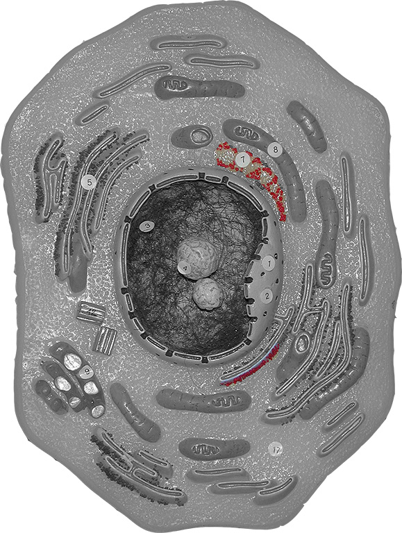
Ribosomes are the exception to the membrane-bound criterion for an organelle. They are considered organelles but are not surrounded by a membrane. Recall ribosomes are made in the nucleoli of the nucleus. They migrate into the cytoplasm after formation. They are suspended in the cytoplasm as small and large subunits. When a messenger RNA is present they join together and attach to the mRNA as a unit.They 'read' the mRNA molecule and produce proteins using the mRNA template. Some ribosomes are associated with the endoplasmic reticulum (another organelle). The area of the endoplasmic reticulum (ER) that has ribosomes attached to it is called rough endoplasmic reticulum. Ribosomes attached to the ER are shown as red dots attached to the membrane to the lower right side of the nucleus. Free ribosomes are shown in red to right above the nucleus.
|
|
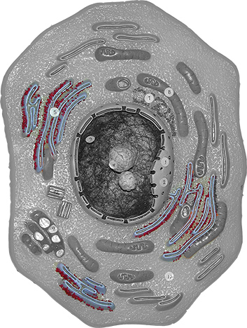
The endoplasmic reticulum (ER) is the most extensive membrane system in cells. In some cells the ER makes up over 40 % of the cells internal membrane structures. The ER in continuous with the nuclear membrane, stretches throughout the cytoplasm and merges with the plasma membrane. Because of this, the ER is thought to be a major transport mechanism in the cell. Molecules could literally leave the nucleus and travel through the ER to the outside environment. The ER consists of membranous hollow tubules. The inside of the tubules is called the cistern (cisternae pl.). The tubules have pores that allow proteins produced by ribosomes on the surface of the ER to move into the cisternae and to be transported out of the cell.
Microscopically, the ER can appear either as a smooth membrane or as a bumpy, rough membrane. The rough membrane is called rough endoplasmic reticulum. It appears rough because ribosomes are attached to it. Ribosomes can attach and detach at will from the surface of the ER. In the image to the right the rough ER is indicated as the blue, flat membrane structures. The red dots on the surface of the membranes are the ribosomes. The smooth ER is present but not color delineated.
The rough ER is responsible for protein production. Cells, like the pancreatic acini cells that produce digestive enzymes which are secreted into the intestine contain large amounts of rough endoplasmic reticulum (RER). The smooth endoplasmic reticulum is associated with cells that have high lipid production/metabolism, steroidal hormone production and are active in detoxification.
|
|
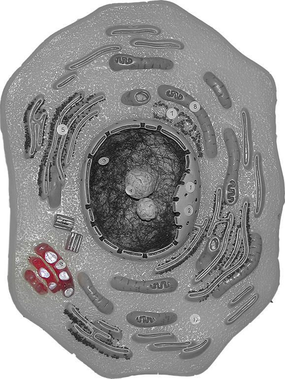
The Golgi Apparatus is shown in red in the image located to the right.In electron micrographs it looks like a stack of pancakes. The Golgi is a stacked membrane organelle. The Golgi is like the post office of the cell. It takes packages produced by other organelles like the ER and stamps and redirects them to the appropriate cellular location. The cell is a big place to a molecule. After a molecule is produced, how does it know where to go? Is a nuclear protein or a plasma membrane protein? You don't want to stick the wrong protein in a membrane! The Golgi figures all of that out. If a protein is meant for the plasma membrane the Golgi wraps it up in a vesicle and stamps it (adds a marker) and sends it off to the plasma membrane where the protein gets incorporated. The Golgi also packages secretory products. So when cells want to dump product outside of the membrane they use the Golgi Apparatus in exopinocytosis. The Golgi also produces vesicles that become lysosomes.
Lysosomes are small membrane-bound vesicles produced by the Golgi Apparatus that contain digestive enzymes (lysozyme, acid hydrolases). These digestive enzymes are important in cell recycling and in cell immunity/protection. Organelles wear out just like your appliances at home will eventually wear out. Cells however are not throw-away societies. When a mitochondrion wears out, a cell can't leave it by the curb and just pop over to Walmart and pick up another. Instead the cell lysosomes will fuse with the defunct organelle and break it down to its components. Those components are released back into the cell to be reused or are discarded outside the plasma membrane. Similarly, some body cells are phagocytes, they eat other cells. When a white blood cell (phagocyte) eats a bacterium it encloses it in a vesicle. Lysosomes fuse to this vesicle, dump in their digestive enzymes and destroy the bacterium.
Peroxisomes are small membrane-bound vesicles that contain 'oxidase' enzymes. These enzymes are involved in detoxifying toxic compounds including various oxygen species and in the breakdown of long chain fatty acids. Tissues like liver that are responsible for detoxifying compounds circulating in the blood are rich in peroxisomes.
To view an interactive model of the cell, follow the link below. Select animal cell. If you click on an organelle below the interaction you can view more information on that organelle.
http://www.cellsalive.com/cells/cell_model.htm
|
|
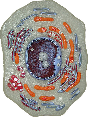
The mitochondrion (mitochondria, pl.) is often called the power-house of the cell. It is the cell organelle responsible for producing the great majority of ATP used by the cell. The mitochondrion is an oblong, cigar-shaped organelle. It has a double membrane structure. The inner membrane, called the cristae membrane is highly folded. This membrane is packed with the components of the electron transport chain. Energy derived from the bonds of organic molecules, notably glucose, is converted into stored energy in ATP by these electron transport chain components.
Interestingly, the mitochondrion is thought to have evolved from a bacterium that invaded another cell. This symbiosis eventually led to the mitochondrion we know about today. The mitochondrion has its own circular DNA chromosome, just like bacteria. The mitochondrion also has its own ribosomes which are identical to bacterial ribosomes, not eukaryotic cytoplasmic ribosomes. Because mitochondria have their own genetic information they have the ability to self replicate. They replicate to meet the energy needs of the tissue. So tissues that are metabolically active will have more mitochondria than tissues that are not metabolically active. Muscles cells will have more mitochondria than fat cells. Muscle cells of athletes will have more mitochondria than muscles of couch potatoes.
The mitochondria are highlighted in orange in the diagram to the right.

All eukaryotic cells have an internal scaffolding called a cytoskeleton. The cytoskeleton gives the cells its shape, participates in whole cell movement and the internal movement of organelles and structures. The cytoskeleton is made up of various elements including microtubules, intermediate filaments and micro filaments.
Microtubules are large (on a microscopic scale) molecules composed of thousands of units of the protein tubulin. Microtubules play an important role in cell division. They make up the spindle apparatus which attaches to chromosomes and moves the chromosomes and sister chromatids around during cell division. Microtubules are also responsible for transport of vesicles within cells. Neurotransmitters are produced by the Nissl bodies located in the soma of neurons but released from the axons of the cell. The cell body and the axon can be pretty far apart. Microtubules extend from the soma to the tips of the axon. Vesicles containing neurotransmitters are shuttled down the microtubules from the cell body to the axon.
Intermediate filaments are fibrous protein molecules that are intermediate in size between the microfilament and the microtubules. Intermediate filaments also play a role in cell shape. They are found beneath the nuclear envelop and determine the shape of the nucleus. They serve as anchors for organelles holding them in place. You may notice in some epithelial tissues that the nucleus is always in the same place, center for simple squamous for example.... the intermediate filaments are holding the nuclei in place.
Microfilaments are the smallest diameter cytoskeletal element. Actin, a contractile globular protein, is the primary protein associated with microfilaments. Microfilaments play a role during cell division also. At the end of nuclear division the cytoplasm must also divide. Microfilaments in the cytoskeleton form the cleavage furrow which allows the cells to 'pinch off' and separate. Microfilaments are also involved in amoeboid movement and the formation of pseudopods. This type of movement is seen in some white blood cells and allows these cells to leave the peripheral blood stream and enter the tissue.
|
|
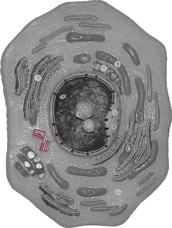
Centrioles are barrel-shaped bodies located near the nucleus in an area called the centrosome in animal cells. They are composed of microtubules. At the onset of mitosis the centrioles duplicate and move to opposite sides of the cell. The centrioles, sometimes called asters are the origination point for the microtubules that will attach to and move chromosomes during cell division. The microtubules grow away from the centrioles producing a star-like network, hence the name asters.
Centrioles also form the basal body of cilia and flagella which are important in movement.
The centrioles are highlighted in red in the photograph to the right.

Musical rendition of the cell:

One of the primary characteristics of all living things is their ability to reproduce. All living things reproduce. Reproduction occurs at both the somatic cellular level and at the organismic level. Meiosis and the generation of germ cells (ovum and sperm) will not be discussed this semester. Mitosis which is cellular or somatic division is one of the topics of today's lab. Mitosis is the process by which a typical body cell replicates to produce two identical offspring. This is an essential process for growth and repair of tissues. It is a process which occurs millions of times a day in every human being. Estimates are that approximately 8,000 new blood cells are produced in your body every second. That is a lot of cell division!
Cell division or mitosis is actually characterized by two separate events, division of the nuclear material (mitosis or karyokinesis) and division of the cytoplasm (cytokinesis). Inherent in our understanding of cell division is the assumption that our daughter cells will be viable, living cells. For our daughter cells to survive they must have all of the master instructions (DNA) needed for life and an adequate supply of raw materials (cytoplasm) to enable metabolism. I liken cell division to building a house. In order to build a house you need a detailed blueprint (DNA) and the blocks, wood and morter (cytoplasm) with which to build the structure. You can't build a house without blueprints and you can't build a house without raw materials. Similarly cells can't survive without all of their 'instructions' or without their raw materials.
The cell cycle describes the events that occur in a cell from one point in cell division until that cell reaches that same point in cell division. It does not describe the birth, life and death of a cell as you would see in a typical life cycle.The cell cycle is divided into two major phases, interphase and mitosis. Cells spend the majority of their time in interphase. Some cells, once differentiated never divide again. They continue to metabolize and function but do not divide. Cells in this non-divisional state are said to be in G0. Cell signals tell the cell when to begin the process of active cell division. Understanding these cell signals is key to turning cell division on and off and may hold the key to treating cancer, paralysis, blindness, etc.
Interphase is subdivided into three other phases: G1, S and G2. During the early days of cell biology, scientists had a difficult time understanding the cell process and what was going on in the cell. G1, stands for the first gap. Scientists knew something was going on, they just weren't sure what it was. Today we know that cells in G1 are making everything they need for 2 cells, more protein, more organelles, etc. The next phase is the S phase or the synthesis phase. During S the cell replicates its DNA. So in human cells, a skin cell for example, at the completion of the S phase the cell will contain 92 chromosomes. Once the DNA has replicated the cell has made the commitment to continue through cell division. There is no turning back. The chromosomes are still present as chromatin. DNA is not visible as chromosomes. G2 is the final subphase of interphase. During G2 the cell continues to grow and produce materials needed for 2 cells. Additionally the cell prepares for mitosis by organizing the machinery needed for cell division like, microtubules.
|
|
|
Now that everything is in place the cell can actually divide. Mitosis is divided into 4 separate phases, prophase, metaphase and anaphase.
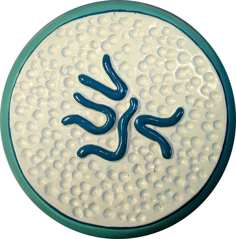
Prophase is the first phase in mitosis. During prophase the chromatin condenses, ie. it coils and packs together to form visible structures. The nucleoli disappear. The nuclear envelop dissolves. The centrioles divide and begin migrating around the cell until they reach opposite polls. Microtubules grow from the centrioles (asters) toward the chromosomes and attach to the chromatids at the centromere region.
The first image to the near right is a photograph of a model of an animal cell during prophase. The photomicrograph to the far right is of onion root tip. Notice how in prophase the chromatin is very granular to stringy in appearance. There are no nucleoli and there is no nuclear envelop present.
|
|
|
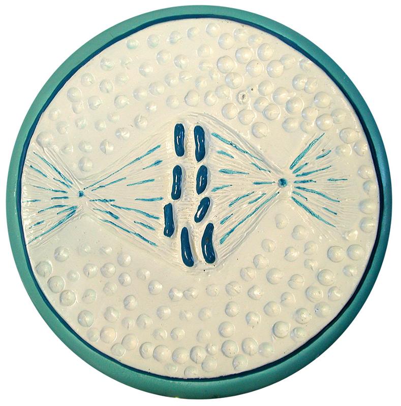
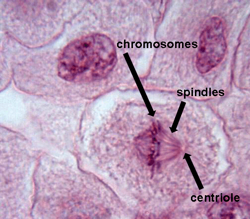
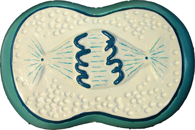
Once the microtubules attach to the chromosomes they start pushing and pulling the chromatid pairs around the cell until they align at the cell equator (midpoint). When this happens the cell is at metaphase.
The first image to the near right is a photograph of a model of an animal cell during metaphase. The photomicrograph to the far right is of whitefish blastula cells. Notice how in metaphase the chromosomes align at the equator of the cell. The chromosomes are the darkly staining bodies. The spindle array is evident in both images.
|
|
|
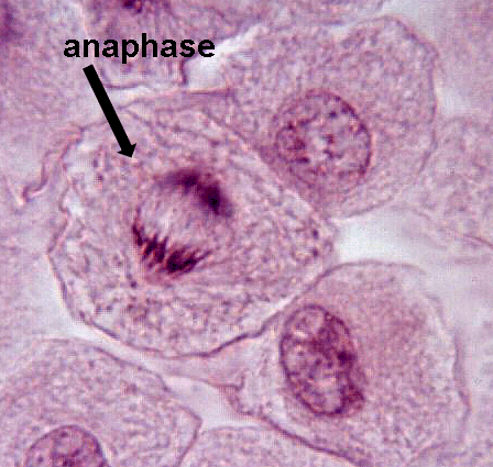
During anaphase the microtubules appear to shorten and pull the sister chromatids apart. As soon as separation is seen between the sister chromatids the cell is in anaphase. Anaphase is the shortest phase of mitosis.
The first image to the near right is a photograph of a model of an animal cell during anaphase. The photomicrograph to the far right is of whitefish blastula cells. Notice how the sister chromatids have separated and a gap appears at the equator. The chromosomes are the darkly staining bodies.
|
|
|
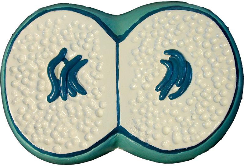
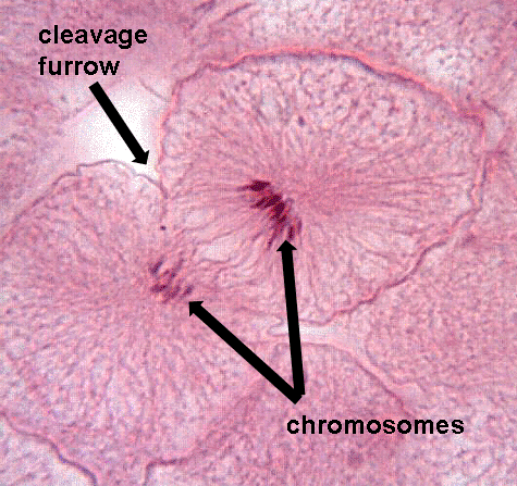
Telophase is the final phase of mitosis. It begins when the sister chromatids reach the poles of the cell and stop moving. Everything that has happened up until this point is reversed. The nuclear membrane reforms; the nucleoli reform; the spindle apparatus dissolves; the chromosomes unwind. At the completion of telophase, the cell contains two nuclei.
The first image to the near right is a photograph of a model of an animal cell during telophase. Notice how the chromosomes are clustered together. A cleavage furrow is evident and the membrane separating the two daughter cells has formed. The photomicrograph to the far right is of whitefish blastula cells. The chromosomes are the darkly staining bodies. They are less distinct.

Cytokinesis is the process by which cells divide their cytoplasm to accomplish the division of the cells. In animal cells this is accomplished through the actions of the cell's cytoskeleton and the formation of a cleavage furrow. Microfilaments align along the equator of the cell. Microfilaments, remember, are composed of actin, a contractile protein. These filaments overlap each other and then start to pull the membrane inward. Since membranes are self-sealing once the membrane is pulled in and touches the membrane on the opposite side of the cell the two daughter cells separate.