

BIOL1611L Organ Systems Exercise
Rat, Torso, Blood, Adipose
Anatomy and physiolgy are disciplines which integrate the structure and functions of organisms from the cellular level to the organismic level. Today, you are going to be introduced to the typical structure of a mammal in the form of the common lab rat. You will be asked to dissect your rat and identify and name specific organs. Additionally, we will start our study of tissues, or histology by learning a little bit about connective tissues and by microscopic examination of slides of adipose (fat) tissue and blood.
All living things exhibit complexity not seen in the non-living world.The cell is the fundamental unit of all life. Cells with a specific function can form a tissue. So cardiac muscle fibers (cells) can work together to form cardiac muscle tissue. Different tissues can work together to form organs. So cardiac muscle tissue, connective tissue and nervous tissue interact to for a heart. A combination of organs with a specific function for an organ system. The tongue, teeth, esophagus, stomach, small intestine and large intestine together form the digestive system. There are 11 commonly sited systems. They are described briefly below.
|
Organ System |
Components |
Functions |
|
Cardiovascular |
heart, blood vessels, blood |
Transport nutrients, protection (immune interactions) |
|
Digestive |
teeth, tongue, glands, esophagus, stomach, intestine |
Physically and chemically breakdown food;absorption of nutrients. |
|
Endocrine |
pancreas, adrenal, thyroid, pituitary, ovary, testis, parathyroid, pineal, thymus |
Production of hormones that assist with maintenance of homeostasis |
|
Integumentary |
skin, cutaneous sense organs and glands |
Protection, excretion, regulates body temperature, vitamin D production |
|
Lymphatic/Immune |
spleen, lymph nodes, thymus, tonsils, diffuse lymphatic tissue |
Protects the body from invasive pathogens and rogue body cells, recirculates fluid from circulatory system. |
|
Muscular |
muscles |
Moves the body, substances through the body, heat production |
|
Nervous |
Sensory cells, special senses, nerves, spinal cord, brain |
Interprets internal and external stimuli; aids in maintaining homeostasis |
|
Respiratory |
lungs, trachea, pharynx, larynx, nasal cavity |
Allows for the exchange of gases; assists with carbonate/bicarbonate balance. |
|
Reproductive |
ovary, testis, oviducts, uterus, vagina, prostate, penis, scrotum, 'plumbing' and accessory glands |
Produces germ cells necessary for the propagation of the species. Produces hormones that produce secondary sexual characteristics. |
|
Skeletal |
bones, tendons, ligaments |
Supports and protects the body and organs.Blood cell formation, calcium storage. Along with the muscles assists with movement of the body. |
|
Urinary/excretory |
kidneys, bladder, urethra, ureters |
Removes metabolic toxins. Maintains water balance of the body. |
To dissect,literally means to expose to view. It is important when doing dissections to make sure you know what you are doing before you start cutting. You do not want to hack and chop. You want to view all of the structures in place or in situ, so work carefully with your specimen. Scissors, not the scalpel are the preferred instrument particulary when you are cutting through the skin and body wall. Lift the structures away from the underlying tissues and organs with your fingers and make a small incision with your scissors. Then insert the sharp point of the scissor into the opening and cut as you lift the tissue (skin or abdominal muscle layer).
Follow the directions given in your laboratory manual.
1. Expose the abdominal muscles by first cutting through the skin. Peel the skin back and pin the skin down to your pan.
2. Carefully cut through the abdominal muscles along the midsagittal line. You can do this by lifting the muscle with forceps (tweezers) and inserting your scissors above the organs.
3. You will need to cut through the ribcage.
4. Make tranverse cuts below the diaphragm to allow you to open these cavities easily.
Identify the organs in the rat's thoracic and abdominal cavities. Use your lab manual as a guide. Some of the organs are shown in the photographs below.
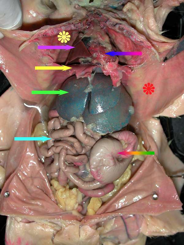 The diaphragm is shown at the tip of the yellow arrow. The space superior to the diaphragm is the thoracic cavity.
The solid pink arrow is pointing to a lobe of the right lung.
The yellow flower is located on the serous membrane surround the pleural cavity, ie. parietal pleura.
The blue to red rainbow arrow is touching the parietal pericardium, the sac surrounding the heart.
The thymus is visible superior to the heart in the mediastinum.
The red flower is on the serous membrane of the abdominal wall or the parietal peritoneum.
The light green arrow is pointing to a lobe of the liver, the largest abdominal organ. Notice the shiny surface of the liver and the other organs. They are shiny because the organs are encased in serous membranes called visceral serosa.
The light blue arrow is pointing to the small intestine.
The red to green rainbow arrow is pointing to the cecum of the large intestine.
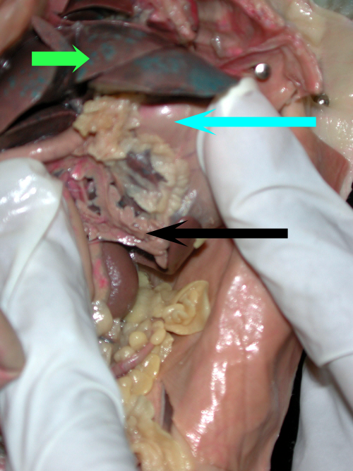
The pancreas is an essential gland with both endocrine and exocrine functions. The pancreas is located in the first curve of the small intestine (duodenal region) where it attaches to the stomach. To see the pancreas you will need to lift the stomach up and away to locate the mesentary in which the pancreas is located. The pancreas is the roast-beefy-looking gland at the tip of the black arrow. The light blue arrow is the stomach. The ribbon of fat along the superior curve of the stomach (starting above the black arrow head) is the lesser omentum. The pale pink tube under the person's left thumb is the small intestine or duodenum. The bright green arrow is pointed at the liver.
Continue with the dissection as outlined in your laboratory manual. Find and identify the components of the reproductive and urinary tracts. Find the esophagus and trachea.
Examine the human torso models in lab and make sure you can identify the organs, cavities, quadrants and regions.
Additionally, in this lab you will start your exploration of histology by learning about two of the connective tissues, blood and adipose tissue. The presentation below is the pre-lab presentation for this laboratory exercise and discusses connective tissue.
